Apoptosis, Necrosis, and NETosis: The Three Engines of Cell-Free DNA
Not all DNA in the blood is the same. We break down the three biological mechanisms that release DNA—programmed death, traumatic injury, and immune defense—and what they tell us about a patient's disease.
Apoptosis, Necrosis, and NETosis: The Three Engines of Cell-Free DNA
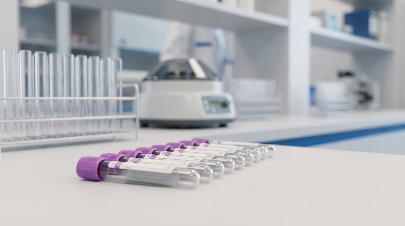
When we receive a laboratory report stating that a patient has "elevated cell-free DNA (cfDNA)," it is easy to view it as a single, monolithic number. But biologically, that number is a sum of many parts.
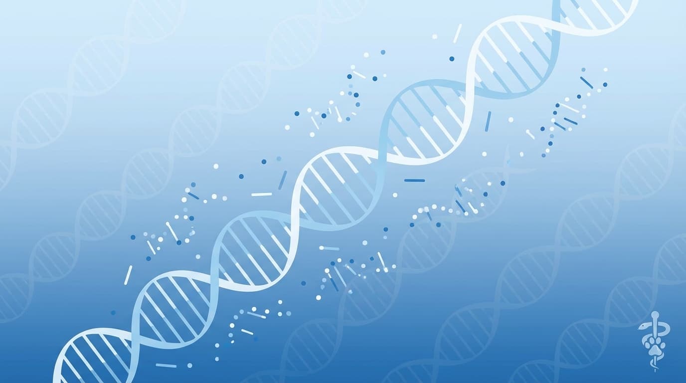
DNA does not simply leak out of healthy, intact cells. It must be released. The mechanism of that release dictates the structure of the DNA fragments and gives us vital clues about the underlying pathology. In veterinary medicine, we are primarily concerned with three release engines: Apoptosis, Necrosis, and NETosis.
1. Apoptosis: The Silent Turnover
Apoptosis is "programmed cell death." It is the body's tidy, highly regulated method of recycling old or damaged cells. In a healthy dog, billions of hematopoietic cells (mostly white blood cells) and epithelial cells (gut lining) undergo apoptosis daily.
The Mechanism
During apoptosis, the cell systematically dismantles itself. Specialized enzymes called caspases activate endonucleases, which cut the genomic DNA at precise intervals. Specifically, they cut the "linker DNA" between nucleosomes (the protein spools DNA wraps around).The Signature: The "Ladder"
Because the DNA wrapped around the nucleosome (~147 base pairs) is protected, apoptotic DNA appears in circulation as a distinct peak at ~167 base pairs (bp) (nucleosome + linker).* Clinical Relevance: This is the source of "baseline" cfDNA in healthy animals. A moderate elevation in apoptotic DNA might be seen in conditions with high cellular turnover that isn't necessarily destructive, or during the early response to chemotherapy when tumor cells are induced to commit suicide.
2. Necrosis: The Chaotic Explosion
Necrosis is "accidental" or traumatic cell death. It occurs when cells are subjected to extreme stress: ischemia (loss of blood flow), thermal injury (burns/heatstroke), toxins, or mechanical trauma. In oncology, we see necrosis in rapidly growing tumors that outstrip their blood supply, leaving a dead core.
The Mechanism
Unlike apoptosis, necrosis is unregulated. The cell membrane ruptures, and the cellular contents spill out. The DNA is not neatly chopped by enzymes; it is randomly degraded by whatever nucleases happen to be nearby.The Signature: The "Smear"
Necrotic DNA fragments are messy. They are often much larger than apoptotic fragments (thousands of base pairs long) because the meticulous cutting process didn't happen.* Clinical Relevance: High levels of long, necrotic DNA fragments are a hallmark of aggressive malignancy (like hemangiosarcoma), severe trauma (HBC), or ischemic events (GDV, splenic torsion). If you see a high "DNA Integrity Index" (a ratio showing many long fragments), think tissue destruction.
3. NETosis: The Immune Weapon
This is the newest player on the field, and perhaps the most important for critical care. NETosis is a specific type of cell death used by neutrophils.
The Mechanism
When neutrophils encounter massive inflammation or bacteria (sepsis), they can eject their chromatin (DNA) into the extracellular space. This forms a Neutrophil Extracellular Trap (NET)—a sticky web of DNA decorated with antimicrobial enzymes and histones designed to physically trap and kill bacteria.The Signature: The "Sticky Web"
NETs are large, complex structures. They contribute significantly to total cfDNA concentration in inflammatory diseases.* Clinical Relevance: NETosis is a massive driver of cfDNA elevation in Sepsis and Immune-Mediated Hemolytic Anemia (IMHA).
* The Danger: While NETs kill bacteria, they are also pro-thrombotic. The DNA backbone acts as a scaffold for platelet aggregation. This explains why dogs with severe IMHA or sepsis are at such high risk for pulmonary thromboembolism (PTE)—the "DNA webs" in their blood are literally triggering clots.
Summary
* Apoptosis: Neat, small fragments. (Turnover).
* Necrosis: Messy, large fragments. (Trauma/Tumor).
* NETosis: DNA webs. (Inflammation/Clots).
Understanding these origins moves us from simply asking "Is it high?" to asking "Where did it come from?"