The Golden Rules: A Clinic SOP for Preanalytical cfDNA Handling
A missed 10-minute window or a single freeze-thaw cycle can ruin a liquid biopsy. Here is the strict Standard Operating Procedure (SOP) every clinic needs.
The Golden Rules: A Clinic SOP for Preanalytical cfDNA Handling
In veterinary diagnostics, we are accustomed to robust analytes. You can leave a serum tube on the counter for a few hours before running a chemistry panel, and the creatinine value will still be accurate. This is NOT true for cell-free DNA (cfDNA).
cfDNA is a trace analyte. In a healthy dog, the concentration might be as low as 0.5 ng/mL. In contrast, a single white blood cell (WBC) contains vast amounts of genomic DNA. If even a tiny fraction of the WBCs in your blood tube rupture (lyse) after collection, they release "genomic noise" that swamps the biological signal you are trying to measure.
To get a result you can trust, you must follow a strict Standard Operating Procedure (SOP).
1. The Draw: Clean and Quick
Goal: Prevent hemolysis (rupture of red blood cells) and activation of clotting.
* Fast the Patient: Lipemia (milky plasma) interferes with many fluorometric assays used to measure cfDNA. A 4–6 hour fast is ideal.
* Peripheral vs. Jugular: Use the largest vein accessible (usually jugular) to ensure a smooth flow. High vacuum pressure or pulling hard on a syringe plunger creates shear forces that tear cells apart.
* The Tube:
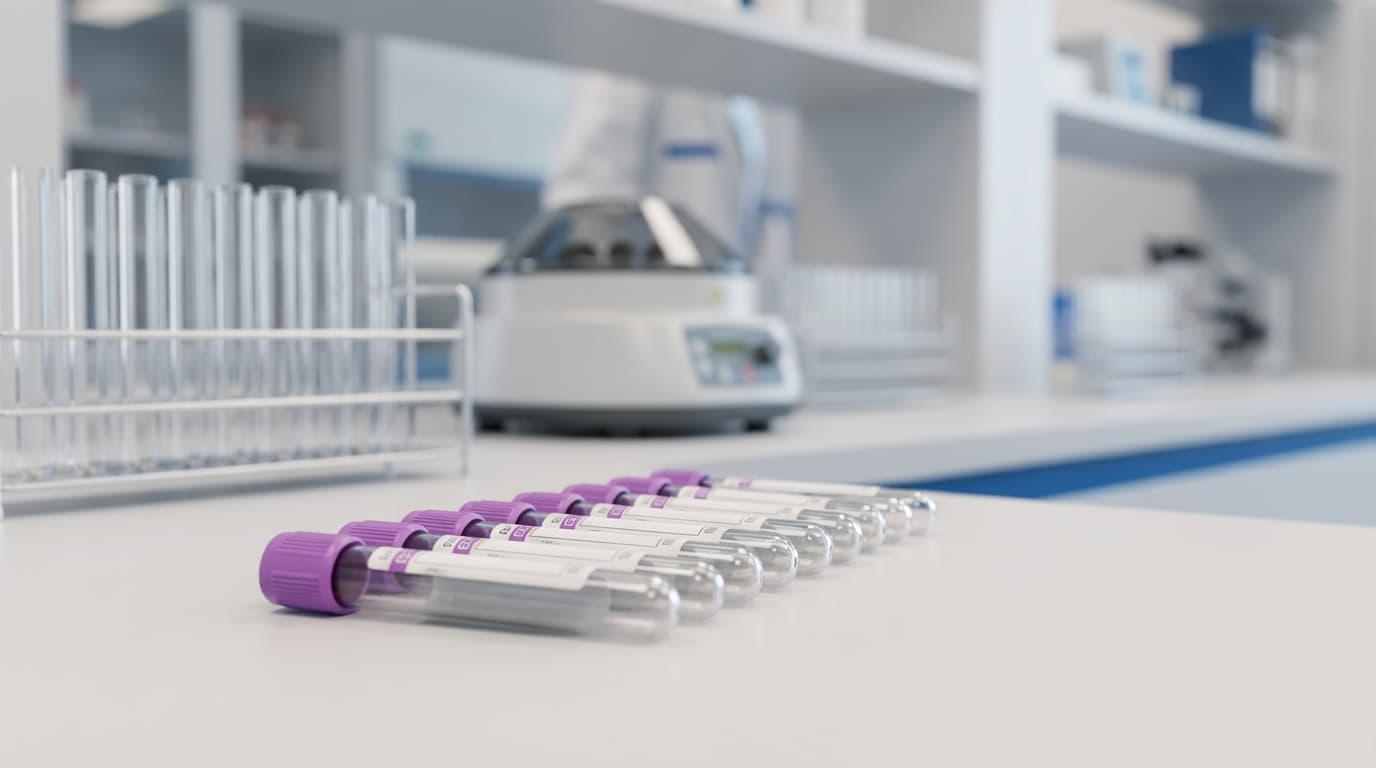
* Standard: K3-EDTA (Purple Top). This is the gold standard for in-house processing.
Forbidden: Serum tubes (Red/Tiger Top). The clotting process requires* cell lysis, which artificially skyrockets cfDNA levels by 200–1000%. Never use serum for liquid biopsy.
Specialty: Cell-Stabilization Tubes (e.g., Streck BCT). Use these only* if you cannot process the sample immediately (see Section 3).
2. The Clock: The 4-Hour Limit
Once the blood is in an EDTA tube, the clock starts. You have a maximum of 4 hours (ideally <2 hours) to separate the plasma from the cells.
* Why? After 4 hours at room temperature, neutrophils begin to die and leak their DNA. This "genomic contamination" can mask the presence of tumor DNA (ctDNA), leading to false negatives in cancer screening.
* Action: Mark the exact time of the draw on the tube label.
3. The Processing: The "Double-Spin" Technique
Simply spinning the blood once (like you would for a biochem panel) is insufficient. A standard spin leaves platelets and cellular debris floating in the plasma. Platelets contain mitochondrial DNA and can interfere with results. You must perform a Double-Spin Protocol.
Step 1: Low-Speed Spin
* Centrifuge whole blood at 1,500 – 2,000 x g for 10 minutes.
* This pellets the Red and White Blood Cells.
Carefully* pipette the supernatant (plasma) into a generic clean tube. Do not disturb the "buffy coat" (the white layer of WBCs between plasma and red cells).
Step 2: High-Speed Spin
* Take that plasma and centrifuge it again at maximum speed (typically >10,000 x g, or "hard spin" on a microcentrifuge) for 10 minutes.
* You will likely see a tiny, invisible-to-white pellet form at the bottom. This is the residual debris.
* Transfer the top portion of the plasma into your final storage tube (cryovial), leaving the bottom ~10% behind to avoid that debris.
4. Storage and Shipping
* Freezing: If you are not testing immediately, freeze the plasma at -80°C (ideal) or -20°C.
The Freeze-Thaw Danger: Repeated freezing and thawing shears DNA fragments. If you anticipate needing to run the test twice, aliquot the plasma into two separate tubes before* freezing.
* Shipping:
* Frozen Plasma: Must ship on Dry Ice. Thawed plasma degrades rapidly.
* Stabilized Whole Blood (Streck): Ship at Room Temperature. Do not freeze whole blood! Freezing lyses all the cells and ruins the sample instantly.
Summary Checklist
1. EDTA Tube (Purple). 2. Process within 2 hours. 3. Double Spin to remove all cells/platelets. 4. No Hemolysis (Pink plasma = Reject). 5. Ship Frozen (Plasma) or Room Temp (Stabilized Whole Blood).Adhering to this SOP is the difference between a diagnostic breakthrough and a costly, confusing lab error.